
二手徕卡 DMiL 倒置显微镜
产品名称: 二手徕卡 DMiL 倒置显微镜
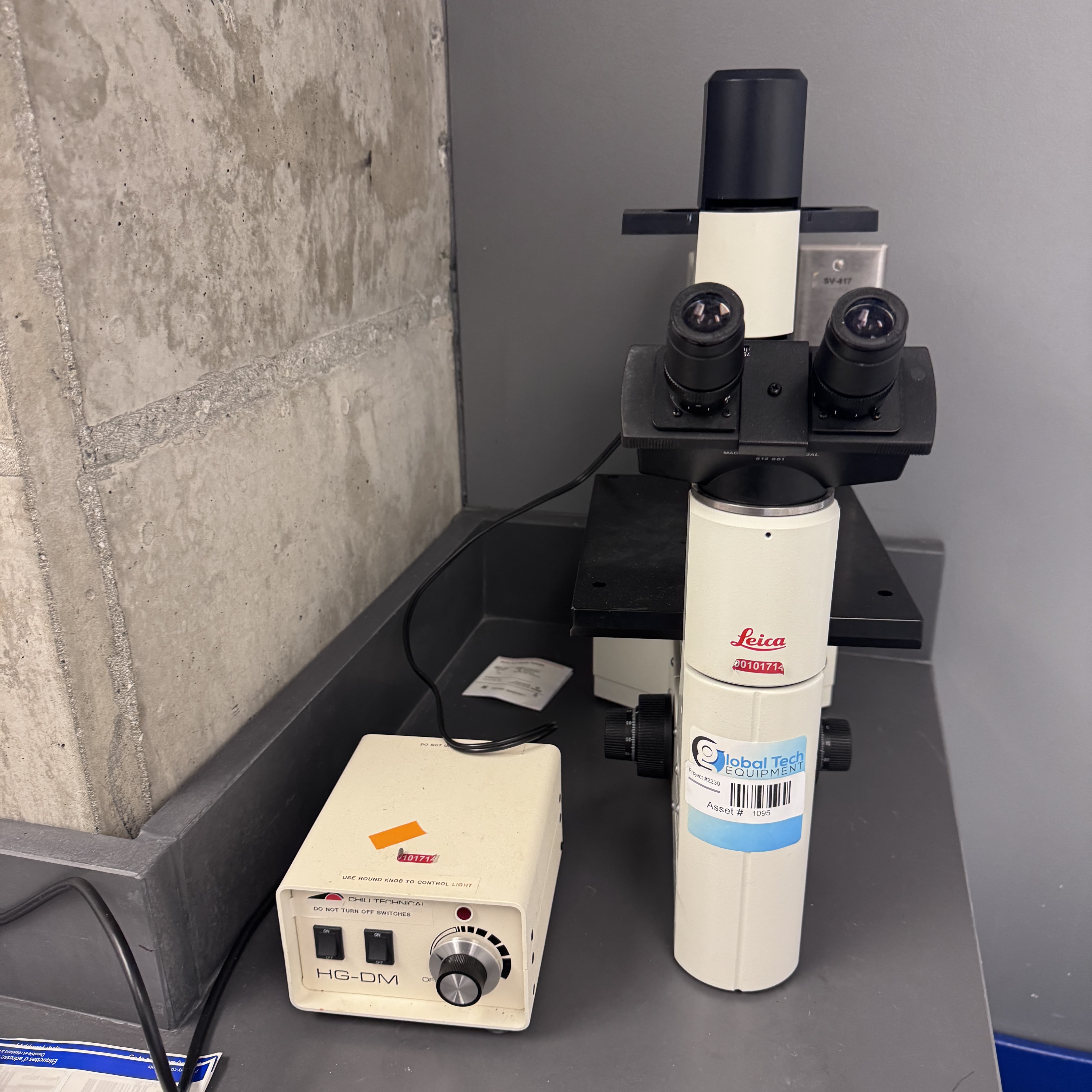
英文名称: Leica DMiL Inverted Microscope
产品编号: Leica DMiL
产品价格: 56900
产品产地: 美国
品牌商标: 徕卡
更新时间: 2026-05-14T16:10:03
使用范围: null
- 联系人 : 陈经理
- 地址 : 上海市浦东新区金桥开发区置业路111号3号楼
- 邮编 : 201299
- 所在区域 : 上海
- 电话 : 137****0200 点击查看
- 传真 : 点击查看
- 邮箱 : sales@yinzhisci.com
- 二维码 : 点击查看
The Leica DMiL Inverted Microscope (often referred to as the DM IL series in its modern LED iteration) is a cornerstone instrument in life science laboratories, specifically engineered for the routine examination of live cell and tissue cultures, microbiology samples, and liquid suspensions. As a product of Leica Microsystems—a brand synonymous with German optical precision—the DMiL combines a robust, space-saving inverted design with high-performance infinity optics to deliver clear, high-contrast images directly from standard culture vessels without the need for slide preparation. Its primary distinction lies in its inverted architecture: unlike upright microscopes where the objective is above the stage, the DMiL positions the objective lenses beneath a fixed or mechanical stage, with the illumination system (light source and condenser) located above. This configuration allows researchers to place bulky containers such as T-25, T-75, or T-175 flasks, 35 mm petri dishes, multi-well plates (6 to 96-well), and even specialized cell factory modules directly onto the stage. The long working distance of the objectives ensures that the lens does not collide with the container bottom, while the ample clearance above the stage provides room for hands-on manipulation, such as pipetting, media changes, or micromanipulation techniques like microinjection and patch-clamp recordings, all while observing the sample through the eyepieces.
Optically, the Leica DMiL is highly versatile, supporting multiple contrast enhancement methods essential for visualizing transparent, unstained living cells. The standard configuration includes Brightfield illumination for observing pigmented or naturally contrasted specimens. For colorless live cells, the DMiL typically employs Phase Contrast (using HI PLAN I or L series objectives with integrated phase rings like Ph1 or Ph2) to convert differences in refractive index into variations in light intensity, rendering cellular structures like nuclei, cytoplasm, and organelles clearly visible. A standout feature of the Leica DMiL (particularly the DM IL LED model) is the inclusion of Leica’s unique Integrated Modulation Contrast (IMC). This technique provides a pseudo-three-dimensional, shadow-cast relief effect similar to Hoffman Modulation Contrast but crucially does not require special objectives; it works with standard plan achromat or plan fluorite objectives. IMC is exceptionally useful for observing subtle morphological changes in cells, monitoring mitosis, checking confluence levels, and inspecting the attachment of cells to the substrate. The illumination system in modern DMiL units is typically a 5W high-intensity LED, which offers a lifespan of up to 50,000 hours, constant color temperature regardless of brightness, and minimal heat emission. This "cold light" is vital for live cell imaging, as it prevents thermal stress on the cultured cells during prolonged observation periods. The LED brightness often auto-adjusts when switching between contrast methods, streamlining the workflow.
For more advanced applications, the DMiL is frequently configured as a fluorescence-capable inverted microscope (sometimes specified as DMiL Fluorescence). It incorporates a fluorescence illumination axis with a manual shutter and a slider that accommodates three filter blocks (e.g., for DAPI, FITC/GFP, and TRITC/mCherry). Some configurations utilize zero pixel shift technology, ensuring that multicolor fluorescent images align perfectly during software overlays. The microscope accepts a binocular or trinocular observation tube; the trinocular port is essential for connecting a digital camera (such as Leica’s ICC series or third-party CMOS/CCD cameras) to the Leica Application Suite (LAS) or LAS X software. This enables image capture, annotation, measurement, and time-live cell imaging. The software, paired with environmental control accessories (like stage top incubators to maintain CO2, temperature, and humidity), transforms the DMiL from a simple inspection tool into a competent live-cell imaging station. The ergonomic design of the DMiL is another strong suit: controls for focusing (coaxial coarse/fine knobs on both sides), condenser height adjustment, illumination intensity, and X-Y stage movement are intuitively placed to minimize user fatigue during long sessions. The stage itself is often a fixed, three-point supported design or a mechanical stage with a removable plate, allowing for quick placement of various vessel holders and reducing the need for constant slide clipping.
The applications of the Leica DMiL Inverted Microscope span the entirety of cell culture-based research and routine diagnostics. In daily cell culture labs, it is the primary workhorse for assessing cell health, monitoring growth and confluence, checking for contamination (microbial or fungal), and verifying the success of passaging or transfection procedures. In pharmacology and drug discovery, it is used for cytotoxicity assays, scratch wound healing assays, and general morphological screening. The instrument is also certified for In Vitro Diagnostics (IVD) in many configurations, making it suitable for clinical laboratories performing IVF (In Vitro Fertilization) procedures or routine pathological sample checks. Its rugged construction ensures stability, while its modularity allows laboratories to start with a basic brightfield/phase model and upgrade to include fluorescence, digital documentation, and advanced contrast methods as their needs evolve. Whether used in a university teaching lab, a biotechnology startup, or a high-throughput screening facility, the Leica DMiL offers a reliable, optically superb, and user-centric solution for the fundamental task of looking at living cells. It strikes an ideal balance between the simplicity required for routine checks and the optical fidelity required for publication-grade documentation, cementing its status as one of the most recognizable and trusted inverted microscopes in the scientific community.
Key Technical Specifications:
|
Parameter |
Specification |
|---|---|
|
Product Name |
Leica DMiL (DM IL) Inverted Microscope |
|
Optical System |
Infinity Color Corrected Optics (ICC) |
|
Observation Tubes |
Binocular (HLT) or Trinocular (for camera) |
|
Objective Revolver |
Typically 4-position, reverse (inward-facing) nosepiece |
|
Standard Objectives |
HI PLAN I (e.g., 4x, 10x Ph1, 20x Ph1, 40x Ph1/L), HI PLAN CY |
|
Contrast Methods |
Brightfield, Phase Contrast (Ph1/Ph2), Integrated Modulation Contrast (IMC) |
|
Illumination (Modern) |
5W LED, 50,000 hour lifespan, constant color temperature, low heat |
|
Condenser |
Long working distance (e.g., S40: 40mm WD, NA 0.45; S80: 80mm WD, NA 0.30) |
|
Stage |
Fixed 3-point support stage or Mechanical XY stage; accepts various inserts |
|
Fluorescence (Optional) |
3-position filter slider; compatible with Hg or LED light sources; DAPI/FITC/TRITC |
|
Camera Integration |
Trinocular port; C-mount adapter; compatible with Leica LAS/LAS X software |
|
Ergonomics |
Coaxial focus knobs (both sides), adjustable tube height, ergonomic controls |
|
Certifications |
Often IVD certified (including IVF use) |
|
Common Vessels |
T-flasks (T-25 to T-175), Petri dishes (35/60/100mm), Multi-well plates, Chamber slides |
|
Applications |
Cell culture inspection, confluence monitoring, live cell manipulation, IVF, microbiology |